Are the brains of men and women differently affected by alcohol and stress?
A new study looks at how the brains of men and women diagnosed with alcohol use disorder respond to stressful stimuli and triggers for alcohol consumption. The differences found are surprising!
Stress is an aggravating factor for alcohol consumption and significantly affects mental health. In this sense, drinking to deal with negative emotions should be considered a warning sign, as it increases the chances of problems with alcohol. In cases where alcohol is already a problem, any stress or trigger, such as seeing images of people drinking, can increase the risk of binge drinking.
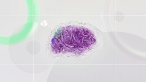
Seeking to understand how biological sex can affect these aspects, researchers at Yale University carried out a survey of 77 participants (46 men and 31 women) diagnosed with disorders associated with alcohol use. These participants were asked to view 3 types of images while “mapping” of the brain activity of these individuals was carried out (a technique called “functional magnetic resonance imaging”). After this mapping, patients were monitored for eight weeks regarding their daily alcohol consumption.
The main objective of the research was to evaluate how different “maps” of brain activity were associated with days of heavy drinking over the following eight weeks. And indeed, the researchers found some significant differences: for example, among women, a lower level of brain activation in a region called the anterior cingulate cortex was found to be correlated with more days of heavy drinking. This finding was not observed in males, who, in turn, had a higher level of activation in brain regions known as the hypothalamus and hippocampus.
All of these regions are important components of the brain reward system, and are linked to drug addiction. Such results lead researchers to point out the possibility that alcohol consumption among female and male people occurs due to the activation of different regions of the reward system, highlighting the need to search for different therapeutic strategies according to the sex of the patient. Although more studies are needed to reach a more solid conclusion on the subject, these results contribute to a general trend in the health sector to provide more individualized therapeutic care, taking into account not only disorders, but various characteristics of the individual, such as gender and age.
References:
Radoman, M., Fogelman, N., Lacadie, C., Seo, D., & Sinha, R. (2024). Neural correlates of stress and alcohol cue-induced alcohol craving and of future heavy drinking: evidence of sex differences. American journal of psychiatry, 181(5), 412-422.
A study evaluated whether it is possible for the thickness of the cerebral cortex of alcoholics to recover to levels comparable to those of people who do not consume alcoholic beverages.
How Alcohol Alters the Brain Cells That Command Our Mind
For decades, it was believed that the brain was composed almost exclusively of neurons, while other cells (glial cells) were seen merely as support. However, recent studies reveal that astrocytes—star-shaped cells—are in fact the active conductors of brain communication, controlling plasticity and the development of the mind.¹ These cells play a decisive role in mental health and are severely harmed by alcohol consumption at all stages of life.
The vulnerability of astrocytes begins very early, since during pregnancy exposure to alcohol interferes with the maturation of these cells.² This process compromises the proper formation of synapses and the blood–brain barrier, directly contributing to the cognitive deficits observed in Fetal Alcohol Syndrome (FAS). During adolescence,³ the impact continues, with alcohol reducing the physical proximity between the processes of these cells and neurons, which impairs the efficiency of connections in the hippocampus—a region vital for the formation of new memories and learning—and in the prefrontal cortex, which acts as the command center for decision-making and impulse control.
In adults with alcohol dependence, astrocytes undergo a profound change known as reactivity, altering their molecular structure and beginning to produce inflammatory substances.² One of the most striking effects of this alteration occurs in the extracellular matrix, where these cells create networks that surround neurons.⁴ Excessive alcohol consumption can make these networks too rigid, trapping neural circuits and promoting behavioral inflexibility and compulsive drinking.
Despite this damage, science is beginning to see new hope for future treatments in these cells. Research in animals indicates that manipulating specific genes within astrocytes—such as through the use of PPAR-γ receptors⁵—can protect memory and learning against the toxic effects of ethanol. Understanding how these cells communicate with the rest of the brain may therefore represent a new frontier for restoring functions lost to alcoholism.
The consequences of this cellular dysregulation extend beyond microscopic biology, manifesting as profound damage to overall health and the integrity of human behavior. Gestational exposure to alcohol is the most common and preventable cause of intellectual disability worldwide, resulting in permanent structural abnormalities and lifelong impairments in cognition and social adaptability. In adulthood, the reduction in the number and density of these cells helps explain the cognitive decline and loss of brain volume observed in individuals with alcohol use disorders. Understanding this relationship is essential, as it suggests that the future of alcoholism treatment may lie in the restoration of these cells.
References